
Heart Anatomy chambers, valves and vessels Anatomy & Physiology
December 7, 2023 - 03:07 am There is a printable worksheet available for download here so you can take the quiz with pen and paper. From the quiz author Ummmmmmm . . . it's pretty self explanatory . . . you label the heart. Just remember one thing - you're looking at the heart like it's in someone else so right and left are switched around.

10+ Explain The Structure Of Heart With Diagram Robhosking Diagram
Coronary circulation Great vessels of the heart Clinical notes Sources Related articles + Show all Heart anatomy The heart has five surfaces: base (posterior), diaphragmatic (inferior), sternocostal (anterior), and left and right pulmonary surfaces. It also has several margins: right, left, superior, and inferior:

Heart Anatomy Anatomy and Physiology II
Labeled heart diagrams Take a look at our labeled heart diagrams (see below) to get an overview of all of the parts of the heart. Once you're feeling confident, you can test yourself using the unlabeled diagrams of the parts of the heart below. Labeled heart diagram showing the heart from anterior Unlabeled heart diagrams (free download!)

Cardiovascular Disease
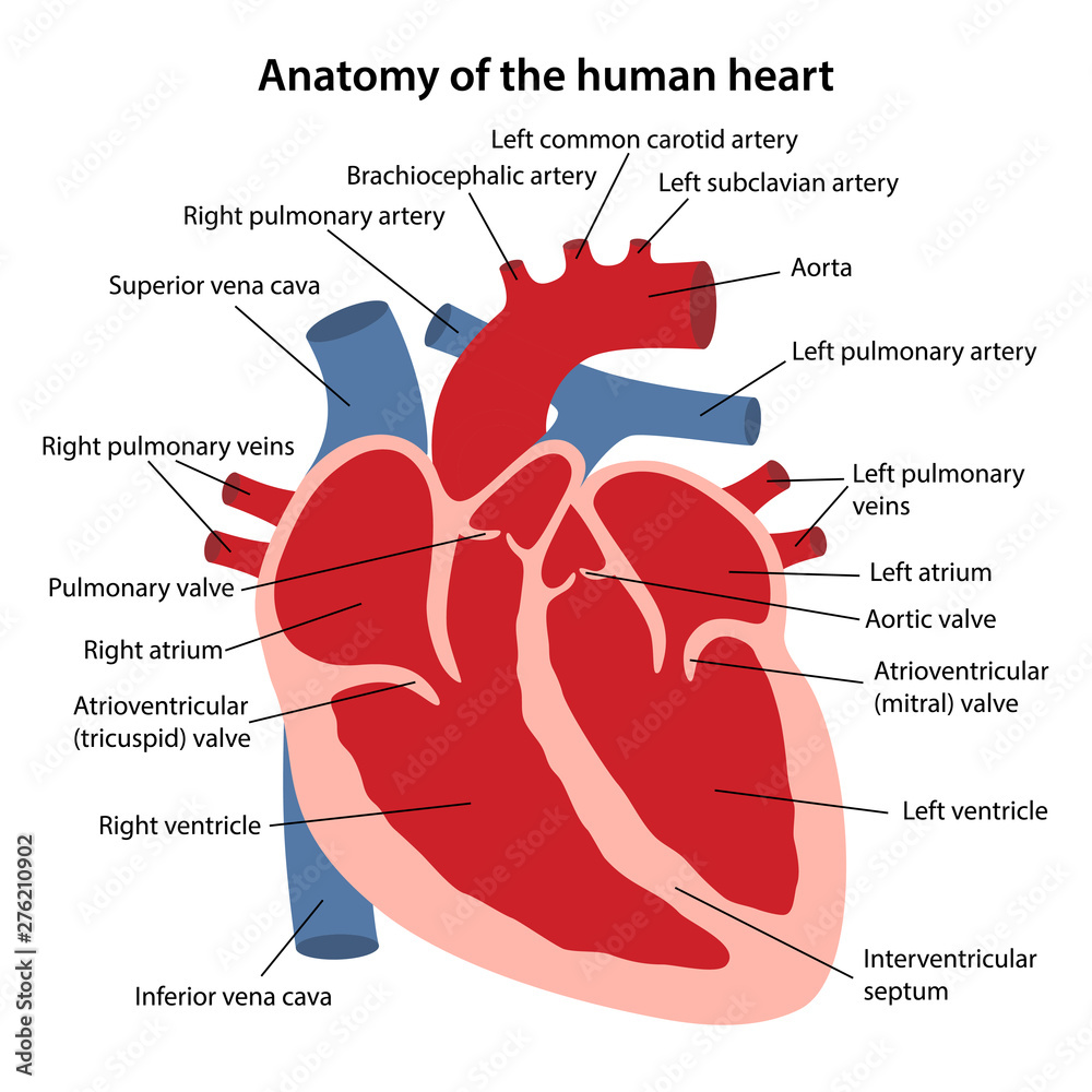
Biology Biology Article Diagram Of Heart Diagram of Heart The human heart is the most crucial organ of the human body. It pumps blood from the heart to different parts of the body and back to the heart. The most common heart attack symptoms or warning signs are chest pain, breathlessness, nausea, sweating etc.
Heart Diagram Labeled
Your heart is the primary organ of your circulatory system. It pumps blood throughout your body, controls your heart rate and maintains blood pressure. Your heart is a bit like a house. It has walls, rooms, doors, plumbing and an electrical system. All the parts of your heart work together to keep blood flowing and send nutrients to your other.
When one teaches, two learn. The heart and the circulatory system (DIAGRAMS)
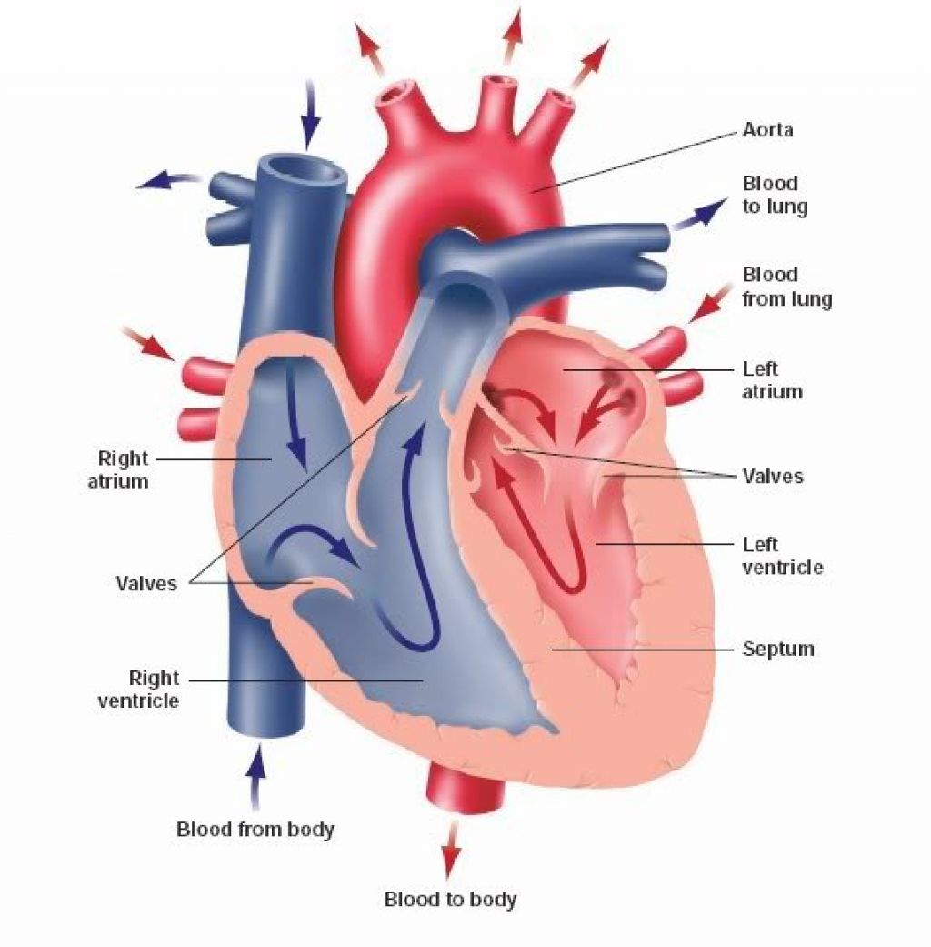
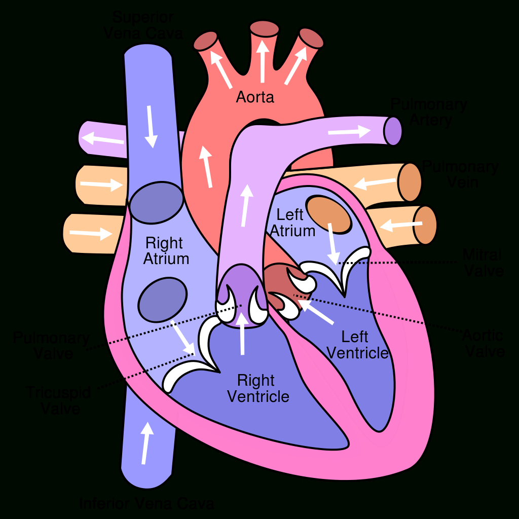
This cycle is then repeated. Every day, the heart pumps about 2,000 gallons (7,600 liters) of blood, beating about 100,000 times. Label the heart anatomy diagram below using the heart glossary. Note: On the diagram, the right side of the heart appears on the left side of the picture (and vice versa) because you are looking at the heart from the.
31 Label The Heart Diagram Label Design Ideas 2020
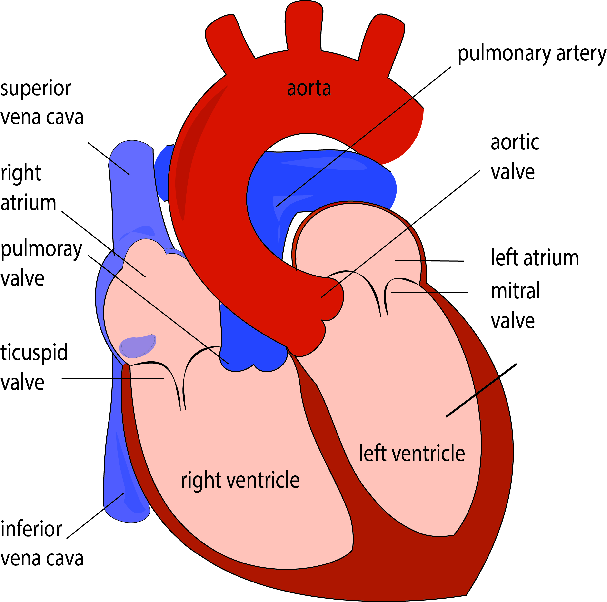
The Heart; The Heart - Map Quiz Game. Aorta; Aortic valve; Left atrium; Left ventricle; Mitral valve; Pulmonary artery; Pulmonary valve; Pulmonary vein; Right atrium; Right ventricle; Septum; Superior vena cava; Tricuspid valve; You need an account to play. Create challenge. 0/0 0 % Game mode: Pin Type Show more game modes. Learn.
Heart Diagram Clipart at GetDrawings Free download
Step 1 and 6 involve a blood vessel, which makes sense as this is how blood enters and exits that side of the heart. Steps 2-5 involve a chamber, valve, chamber, and valve. So if you remember this general pattern, it will help you recall the order in which blood flows through each side of the heart.

Anatomy and Physiology Heart Anatomy
Cardiomyopathy is when the heart muscle becomes enlarged, thick, or rigid. As cardiomyopathy worsens, the heart becomes weaker and is less able to pump blood through the body and maintain a normal electrical rhythm.
.svg/1043px-Diagram_of_the_human_heart_(cropped).svg.png)
FileDiagram of the human heart (cropped).svg Wikipedia
The position of the heart in the torso between the vertebrae and sternum (see Figure 19.2 for the position of the heart within the thorax) allows for individuals to apply an emergency technique known as cardiopulmonary resuscitation (CPR) if the heart of a patient should stop.
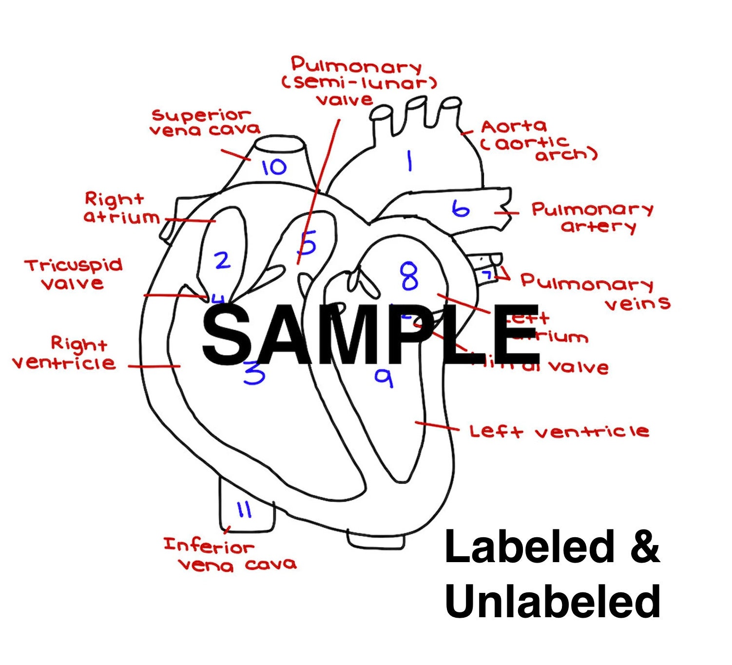
The Heart Diagram Labeled and Unlabeled Worksheets Heart Etsy UK
$9.99 Add To Cart Anatomy of the Heart Welcome to the anatomy of the heart made easy! We will use labeled diagrams and pictures to learn the main cardiac structures and related vascular system. In addition to reviewing the human heart anatomy, we will also discuss the function and order in which blood flows through the heart.
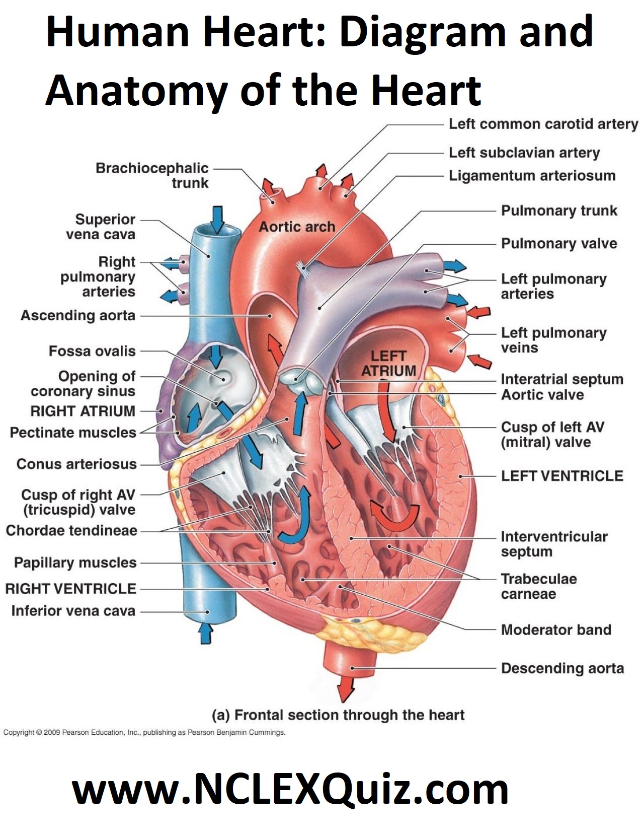
Human Heart Diagram and Anatomy of the Heart StudyPK
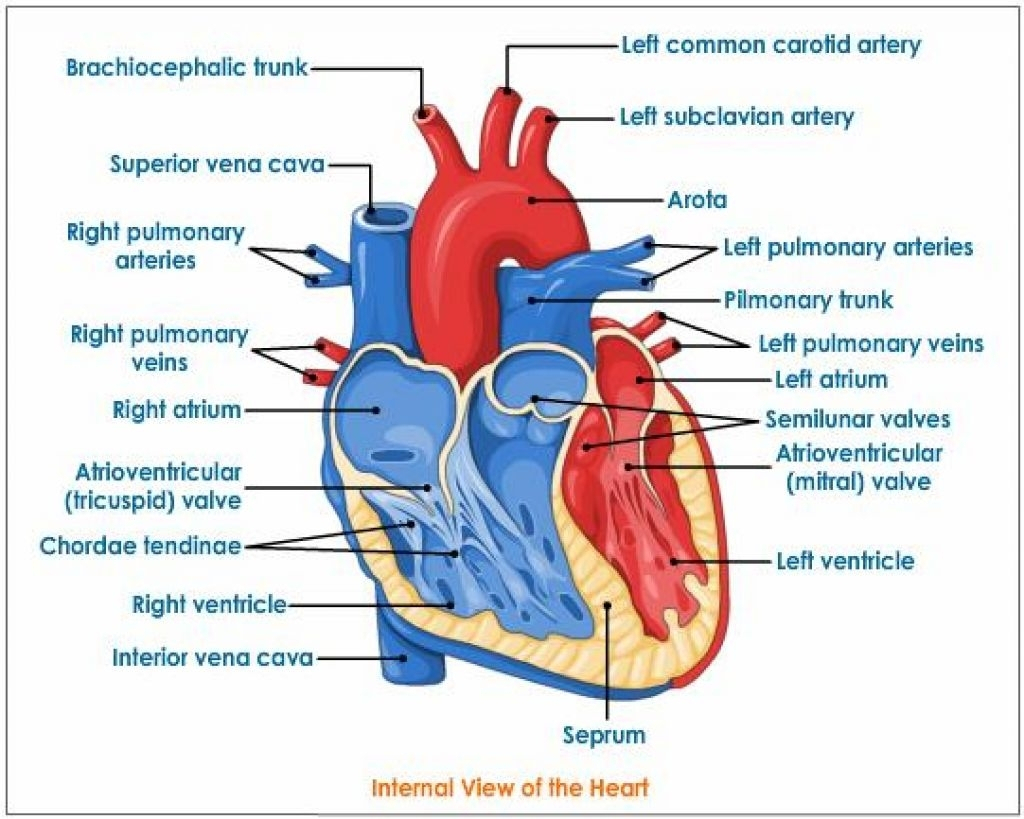
Definition. A vein that is the largest vein in the human body and returns blood to the right atrium of the heart from bodily parts below the diaphragm. + 1 more side. Term. Septum. Definition. Divides the right and left chambers of the heart. + 1 more side. Term.

15 Heart Diagram Labeled Blood Flow Robhosking Diagram
In this lecture, Dr Mike shows the two best ways to draw and label the heart!

Heart anatomy, Heart diagram, Human heart diagram
Labelling the heart. The heart is a muscular organ that pumps blood through the blood vessels of the circulatory system. Blood transports oxygen and nutrients to the body. It is also involved in the removal of metabolic wastes. In this activity, students use online and paper resources to identify and label the main parts of the heart.

Heart Structure Anatomy
The heart is an organ that weighs approximately 350 grams (less than one pound). It's nearly the size of an adult's clenched fist. It's located in the thorax (chest)—between the lungs —and extends downward between the second and fifth intercostal (between the ribs).
On Heart Kardiohirurgija.rs
In this interactive, you can label parts of the human heart. and drop the text labels onto the boxes next to the heart diagram. If you want to redo an answer, click on the box and the answer will go back to the top so you can move it to another box. If you want to check your answers, use the Reset Incorrect button.
